What Is AFP Antibody?

AFP (Alpha-Fetoprotein) is the main plasma protein produced by egg yolk bags, intestinal and liver tracts during fetal life. AFP expression in adults is often associated with hepatoma or teratoma. However, the persistence of AFP hereditary can also be found in individuals without clear pathology.

Protein is considered a serum fetus albumin partner, and AFP gene and albumin are present in the tandem in the same transcription orientation in chromosome 4. AFP is found in the form of monomers, damp, and trimeric. AFP can also tie copper ions, nickel ions, fatty acids and bilirubin. You can also know more about AFP antibodies-online via https://www.bosterbio.com/anti-alpha-1-fetoprotein-picoband-trade-antibody-a00522-boster.html.

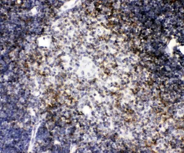
AFP levels in the membranes are used to measure the loss of kidney protein to the screen for spina bifida and anencephaly. A high level of AFP serum has been identified in patients with hepatocellular (HCC) carcinoma (HCC), terroatstastoma, colorectal cancer, pancreatic cancer, and germ cell neoplasms.

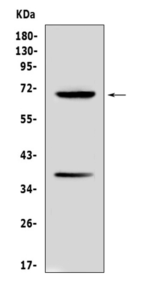
Anti-AFP antibodies are offered by a number of suppliers. This target gene encodes ‘alpha fetoprotein’ protein in humans and can also be known as Alpha-1-Fetoprotein, AFPD, Feta, HPAFP, and Alpha-Feto Globulin. Structurally, proteins reported 68.7 kilodalton in the masses, with subcellular localization issued.

AFP is famous for being the most abundant plasma protein in the human fetus, and to be biomarkers in certain tumors. Alpha-Fetoprotein (AFP) is 65 KDA glycoprotein found in the heart of the mammalian fetus, egg yolk bag, and GI channel. While AFP expression in adult cells is low, it is very stated in adult liver cancer cells.

Know All About Ubiquitin Antibody

Ubiquitin is a conserved polypeptide unit that plays an important role in the ubiquitin-proteasome pathway. Ubiquitin can be covalently attached to many cellular proteins via a ubiquitination process that targets the 26S proteasome-degrading protein. Three components are involved in the conjugation process of the ubiquitin target protein.

Ubiquitin/ UBB antibody is first activated by forming a thiol ester complex with the activating moiety E1; Activated ubiquitin is further transferred to the ubiquitin carrier protein E2, then from E2 to ubiquitin ligase E3 to finally deliver lysine target protein residues to epsilon-NH2.

The ubiquitin-proteasome pathway is involved in a variety of normal biological processes and disease-associated disorders. Several proteins such as IκB, p53, cdc25A and Bcl-2 have been shown to target the ubiquitin-proteasome process as part of the regulation of cell cycle development, differentiation, cellular stress response, and apoptosis.

Ubiquitin, a highly conserved protein that plays a key role in directing cellular proteins to degrade the 26S proteasome, is synthesized as a precursor protein consisting of a single polyubiquitin or ubiquitin chain fused to an unbound protein. This gene encodes a fusion protein consisting of ubiquitin at the N-terminus and ribosomal protein S27a at the C-terminus.

When expressed in yeast, the protein undergoes post-translational processing to produce free ubiquitin monomer and ribosomal protein S27a. The ribosomal protein S27a is part of the 40S subunit of the ribosome and belongs to the family of ribosomal proteins S27AE. It contains the earth zinc domain of C4 and is located in the cytoplasm.


What Is CD4 Antibody?

CD4 is a surface marker of 51 kDa that is expressed on T-cells. Antibodies to CD4 are useful for studying signal transduction. CD4, also known by the T cell antigen T4 (LEU3), is the gene symbol of CD4 molecule. The CD4 antigen plays a role in the recognition and binding of MHC class II molecules. It is also a co-receptor to HIV.

CD4 is expressed mainly in T-lymphocytes, but it may also be expressed in other immune system cells such as macrophages and monocytes. CD4 expression can be found at the tissue level in the thymus and lymph nodes, tonsils and spleen as well as in certain regions of the brain and gut and other non-lymphoid tissues. You can know more about CD4 antibodies online via https://www.bosterbio.com/anti-cd4-picoband-trade-antibody-a00344-2-boster.html.

Through its association with Lck and the T-cell receptor compound, CD4 can initiate or enhance the initial phase of T cell activation. It can also be used to mediate neuronal damage caused by immune-mediated and infectious diseases of the central nervous systems.

CD4 is a 55kDa type 1 membrane glycoprotein. It is found primarily on most thymocytes, and a subset mature T lymphocytes. CD4 is also expressed in humans to a lesser degree on macrophage-related cells and monocytes. The human CD4 cDNA encodes 458 amino acids (aa) precursor proteins, a 371-aa extracellular area containing four immunoglobulin hology domains and a 24 aa Transmembrane domain, and a 38 Aa Cytoplasmic Domain.

The CD4 coreceptor is required to allow T cells to recognize antigens presented by major histocompatibility complicatedes class II. CD4 is a coreceptor for HIV entry. It binds specifically to gp120, an external envelope glycoprotein of HIV.

Know Everything About A Cell Staining Kit

The cell stain kit can be used to determine cell viability prior to intracellular antibody staining, or for the elimination of hazardous substances using formaldehyde fixation. This kit has been optimized and validated for use with a violet laser flow cytometer. Dyes are freeze dried in separate vials to maintain stability.

Staining pattern is the same before and after fixation. Minimal spectral overlap between other fluorophores. The cell staining equipment is packaged in convenient 40-test vials, which are not available in solution. This helps to ensure the dye's stability and performance over time. The effectiveness of amine reactive dyes in solutions will decrease over time.

It is best to use the vial immediately after rehydrating. If it is impossible to do so, you can aliquot vials and keep them at -80C. Fixable cell stain binds covalently with intracellular and extracellular aminos. The staining pattern can be preserved after formaldehyde fixation. 

The excitation limit for the aqua-fluorescent reactive dye is 375 nm. However, it can be excited well by the 405 nm violet light laser. In cells with compromised membranes the dye reacts with both the cell interior and the cell surface to produce intense fluorescent staining. The dye's reactivity in viable cells is limited to cell-surface amines.

This results in less intense fluorescence. It is possible to distinguish between dead and live cells by the difference in intensity, which is usually greater than 50-fold.